


Institute Of Invasive & Non Invasive Radiology
We at Padiyath Medicity- Hopital Du Cinquantenaire committed to provide state of the art high quality diagnostic services to our patients and referring physicians.
Institute Of Invasive & Non Invasive Radiology
We at Padiyath Medicity- Hopital Du Cinquantenaire committed to provide state of the art high quality diagnostic services to our patients and referring physicians. Our facilities are equipped with high end, latest generation machines with cutting edge technology. This coupled with the highly qualified team of Doctors, skilled technicians , nurses and supporting staff provide exceptional patient care and services round the clock.
This Center provides imaging and interpretation services to all In- Patient and Out-Patient facilities. All forms of diagnostic and interventional radiological procedures including sophisticated vascular and neuro interventional procedures are performed.
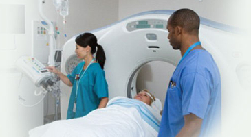
Our Center has several state-of-the-art equipment, more than 60 personnel working for the diagnostic and therapeutic services. The Department provides a wide array of diagnostic imaging tests utilizing the most advanced imaging equipment available ranging from conventional x-ray to 64-slice CT and MRI Scan.
Services Offered
- Magnetic Resonance Imaging(MRI)
- Digital Substraction Angiography (DSA)
- Mammogram
- Ultrasonography
- Barium Studies
- Micturating Cystourethrogram (MCU)
- Interventional vascular Studies
- Multidetector Computerized Tomography (16 and 64 slice)
- Digital X-ray
- Color Doppler
- Special Investigations
- Intravenous Pyelogram (IVP)
- Portable X-ray
Computed and Digital Radiography
Flexible, high quality digital imaging solution enables seamless integration of the X-ray generator with the hospital or radiology information system. Innovative high end advanced flat panel detectors provide outstanding image quality, optimal workflow and operability. Computed Radiography (CR) provides digital x-ray images, while Digital Fluoroscopy allows the digital viewing of deep structures of the body in real time.
Portable x-ray machines, movable around the hospital, are used for convenient x-ray purposes. The portable C-arm x-ray machine is used in OT for instant x-rays of bones and fractures, as well as to ensure that catheters have been placed correctly, to check bile ducts, and to locate pacemakers.
BMD & Colour Doppler
BMD
Evaluation of bone mineral content is done by DEXA method with grading of the severity of osteoporosis by World Health Organization criteria.
Colour Doppler
We have a high end 4D color doppler equipment (Aloka) that has a superior gray scale resolution and picks up even very low velocity flow and the smallest of plaques / turbulent flow velocities.Carotid, Renal, Peripheral vascular ( upper and lower limb), Portal, Scrotal and Obstetric Doppler studies can be performed.
Magnetic Resonance Imaging (MRI)
It is a technique used in Radiology to visualize in detail the internal structures of the body. It provides excellent contrast between the various soft tissues of the body. It is very useful in Imaging in Brain, Spine, Joints, Muscles, Blood Vessels, Chest, Abdominal and Pelvic Organs including the Breast, Heart, Prostate and Ovaries.
We have the State-of-the-art Siemens MRI machine with an open bore magnet. It is one of the most advanced equipments available for the whole body imaging. . The MRI machine in the Padiyath Medicity - Hopital Du Cinquantenaire provides high quality images and is backed by skilled professionals to provide interpretation and reporting services.
Computed Tomography (CT) or Computed Axial Tomography (CAT)
Computed Tomography (CT) or Computed Axial Tomography (CAT) is an imaging method using focussed X-ray beam to generate an image of a body part in an axial plane to acquire a 3-D data. Computerised Tomography utilises X-rays directed from a 360º gantry to provide high resolution multi-planar images. The Siemens computerised tomography machine, available in the Diagnostic Suite, is ideal for advanced orthopaedic, cranial and abdominal imaging.
Digital Subtraction Angiography Lab
Digital Subtraction Angiography Lab
This highly sophisticated equipment provides crystal clear images of abdominal, renal, cerebral and peripheral blood vessels for the diagnosis of patients with gastrointestinal as well as peripheral vascular and neurological problems.
Among the many important procedures and treatments possible with the use of this equipment are balloon dilation and stenting to repair and maintain obstructed vessels (angioplasty), embolizations to eliminate bleeding, chemoembolization and radiofrequency ablation for cancer treatment and advanced procedures to assist liver transplantation. The department has also started offering treatments for stroke (carotid stenting) and for brain vascular problems (AVM embolisations and aneurysm coiling).
Ultrasound system
The premium performance ultrasound system captures crisp, high-resolution images even in technically challenging situations. It makes easy to add 3D imaging to any exam by removing the barriers to volume imaging and a host of workflow enhancers facilitate faster exams, enabling perfect image optimization.
Our technologically advanced ultrasound capabilities have expanded to include vascular, breast and obstetrical imaging with subspecialty interpretation for optimum patient care. Services include:
- Abdomen-limited and complete
- Obstetrics
- Peripheral vascular
- Pelvis
- Thyroid
Flouroscopy
Fluoroscopy is an x-ray (radiography) that makes it possible to see internal organs in motion. It is used to diagnose gastrointestinal (GI) tract disorders. GI procedures require a contrast material called barium. The GI tract is filled with barium, the radiologist is able to view and assess the anatomy and function of the rectum, colon or part of the lower small intestine.
The equipment used for this examination usually consists of a radiographic table, an x-ray tube and a television-like monitor that is located in the examining room. The images are typically captured electronically and stored in a computer called PACS (picture archive communication system).
Digital Mammography
Digital (computerized) mammography is similar to standard mammography in that X-rays are used to produce detailed images of the breast. Digital mammography uses essentially the same mammography system as conventional mammography, but the system is equipped with a digital receptor and a computer instead of a film cassette.
From the patient's perspective, a digital mammogram is the same as a standard film-based mammogram in that breast compression and radiation are necessary to create clear images of the breast. The time needed to position the patient is the same for each method. However, conventional film mammography requires several minutes to develop the film while digital mammography provides the image on the computer monitor in less than a minute after the exposure/data acquisition. Thus, digital mammography provides a shorter exam for the woman and may allow mammography facilities to conduct more mammograms in a day.
Digital mammography can also be manipulated to correct for under- or over-exposure after the exam is completed to help the radiologist more clearly see certain areas of the breast. The technologist can review the image just seconds after the exposure, thus decreasing procedure time.
Interventional Radiology
Interventional radiology is a specialized area of radiology that utilizes imaging in combination with minimally invasive procedures to diagnose and/or treat a variety of conditions. The procedures are generally performed using needles or catheters, and the images (including CT scan X-ray, MRI, andultrasound) are used as a guide, allowing your doctor to perform very precise procedures while minimizing pain, recovery time, and other stress to the body.
Some common interventional radiology procedures include:
- Image-guided needle biopsy such as MRI breast biopsyor ultrasound biopsy
- Cancer treatments such as radiofrequency ablation and chemoembolization
- Treatment of peripheral vascular disease
- Kyphoplasty (treatment for deterioration of the vertebrae)
Institute of Invasive and Non Invasive radiology at Padiyath Medicity-Hopital Du Cinquantenaire plays a very important and significant role in the overall health care delivery system and academic activities of the hospital. It also provides platform for research activities and plans to conduct various educational and research activities in near future.